Biosurf IV
Spatial Organisation & Dynamics of Molecules and Cells at Surfaces
September 20th-21st 2001, EPF-Lausanne, Switzerland
Welcome
Scientific Committee
Patrick Aebischer, Swiss Federal Institute of Technology, EPFL
Gaudenz Danuser, Swiss Federal Institute of Technology, ETHZ
Markus Ehrat, Zeptosens AG, Witterswil
Takao Hanawa, National Research Institute for Materials Science,
Tsukuba, Japan
Heinrich Hofmann, Swiss Federal Institute of Technology, EPFL
Margarethe Hofmann, MAT SEARCH Consulting, Pully
Jeffrey A. Hubbell, Swiss Federal Institute of Technology, ETHZ
Lars Nieba, TOP NANO 21, c/oTEMAS AG, Arbon
R. Geoff Richards, AO Research Institute, Davos
Marcus Textor, Swiss Federal Institute of Technology, ETHZ
Introduction to the abstract by
Marcus Textor, ETH-Zurich and Margarethe Hofmann, EPF-Lausanne
BIOSURF IV is the fourth of a series of conferences with international
participation devoted to topics in biomaterials, biosensor and surface
science research and development. The conference addresses scientists
and engineers in academia and industry interested in recent advances
and exchange of ideas in these fields.
BIOSURF IV will cover the topic of space and time resolution
in the investigation of biomaterials and biosensor performance.
We believe this to be a relevant topic both in basic research that
is oriented towards a better understanding of biochemical and biological
mechanisms of phenomena at biomaterials and biosensor interfaces
as well as in the development of materials and devices that are
designed to induce a desired biological response.
Spatial organization of biomolecules and cells is one of
the main topics to be addressed in this conference. Spatial control
over protein and DNA/RNA interactions with bioaffinity sensor chips
is crucial in the context of developing microarray sensing platforms
in DNA/RNA (genomics), protein (proteomics) and in cell-based (cellomics)
sensor devices. The organization of cells on 2D and 3D substrates
providing particular microenvironments is relevant for areas such
as tissue engineering and cell-based sensing. Micro- and nanofabrication
techniques have provided in the last few years new tools for tailoring
surfaces that permit the precise control of the spatial organization
of bioaffinity reactions, cell interactions and tissue formation
at biosensor and biomedical device surfaces.
One of the frequent drawbacks of conventional cell culture testing
protocols is the lack of direct information on the dynamics of
biological processes, causing ambiguities in the interpretation
of the complex biological phenomena that occur at the interface
between cells and biomaterials and upon the inner and outer leaflets
of cell membranes. New instruments and characterization techniques
have been developed that provide a basis for quantitative microscopy
and imaging of biomaterial and biosensor surfaces with both real-time
and high spatial resolution, applied under biologically relevant
in situ conditions.
 |
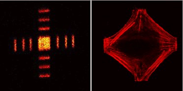
Fluorescence microscopy (right) image depicting a cell (actin fibers labelled with rhodamine appear in red) attached to a cell-adhesive pattern, the ToF-SIMS image of which is shown on the left. Courtesy of R. Michel and J. Lussi (ETH Zurich). See the abstract of Michel et al. for more information. |
